
High burden of germline variants of uncertain significance in advanced renal cell carcinoma: insights from an Indian cohort
Pavan Raju Kola1, Amith Paruchuri1, Nimisha Preman1, Hemavathi Baskarane2, Vishakha Hooda2, Aparna Sharma3, Ishaan Gupta4, Saran Kumar5, Shilpi Minocha5, Pranay Tanwar6, Mohit Kumar Divakar2 and Atul Batra2
1All India Institute of Medical Sciences, New Delhi 110029, India
2Department of Medical Oncology, Dr B.R.A. IRCH, AIIMS, New Delhi 110029, India 3Department of Medical Oncology, National Cancer Institute, AIIMS, Jhajjar, India
4Department of Biochemical Engineering and Biotechnology, Indian Institute of Technology, New Delhi, India
5Kusuma School of Biological Sciences, Indian Institute of Technology, New Delhi, India
6Department of Lab Oncology, Dr B.R.A. IRCH, AIIMS, New Delhi 110029, India
Abstract
Background: Renal cell carcinoma (RCC) is the most common renal malignancy, accounting for nearly 90% of cases. In contrast to targeted genomic profiling, the implementation of whole-exome sequencing substantially broadened the genomic landscape that could be sequenced, leading to the identification of numerous variants that had previously remained uncharacterised, known as variants of uncertain significance (VUS). This study aims to establish a germline VUS-specific database in Indian RCC patients by integrating data from the Indian population and to explore the clinical significance of these variants.
Methods: In this cross-sectional study, whole-exome sequencing was performed in all patients with histologically confirmed metastatic RCC enrolled between September 2023 and August 2024. Variants in RCC susceptibility genes and RCC-related genes were classified according to the American College of Medical Genetics and Genomics guidelines. Variants of uncertain significance were reevaluated using an indigenous population database called IndiGenomes. Descriptive statistics were used to analyse demographic and baseline clinical characteristics.
Results: Among 139 patients included in the study, VUS were identified in 108 (77.7%), with a significantly higher prevalence in females (93%) compared to males (75%) (p = 0.044). Among the 24 genes analysed, 246 VUS mutations were identified in 13 different genes, most frequently in TTN, MUC4 and MUC16. Twenty-nine patients atleast four VUS, with a maximum of 19 variants in a single individual. Importantly, seven patients carried VUS in high-penetrance genes such as BAP1, TSC1 and TSC2, with TSC2 being the most common (n = 5). Of the 246 VUS, 64 variants (26%) were present in the IndiGenomes database, and three variants in MUC4 were reclassified as benign or likely benign.
Conclusion: Patients with advanced RCC have a high prevalence of germline VUS in RCC-associated genes. Given this substantial burden, there is a critical need for comprehensive research aimed at characterising these variants to understand their functional relevance in RCC.
Keywords: variant of uncertain significance, reclassification, renal cell carcinoma, whole-exome sequencing
Correspondence to: Atul Batra and Mohit Kumar Divakar
Email: batraatul85@gmail.com and divakaremt6@gmail.com
Published: 05/11/2026
Received: 13/06/2026
Publication costs for this article were supported by ecancer (UK Charity number 1176307).
Copyright: © the authors; licensee ecancermedicalscience. This is an Open Access article distributed under the terms of the Creative Commons Attribution License (http://creativecommons.org/licenses/by/4.0), which permits unrestricted use, distribution, and reproduction in any medium, provided the original work is properly cited.
Introduction
Kidney cancer remains a significant global health concern, with an estimated 434,840 new cases diagnosed and more than 100,000 deaths reported worldwide in 2022 [1]. Renal cell carcinoma (RCC) accounts for nearly 90% of all renal malignancies and is now recognised as a heterogeneous group of subtypes defined by distinct histological, molecular and clinical features [2,3]. While most cases are sporadic, approximately 5% arise from inherited predisposition syndromes involving high-penetrance genes such as VHL, FLCN, MET, FH, BAP1, SDHD, TSC1, TSC2 and PTEN [4]. Despite the advancements in current research, the American College of Medical Genetics and Genomics (ACMG) referral guidelines for germline testing identify only a subset of patients eligible for the germline testing, which might result in missing a few carriers of pathogenic or likely pathogenic (P/LP) variants [5–8]. This emphasises a shift in germline testing indications towards defining individuals who may not require testing [9–11].
A central challenge in germline testing is the high rate of variants of uncertain significance (VUS). Defined as genetic alterations with unclear clinical impact due to limited evidence and inconsistent interpretation, VUS are reported in RCC at frequencies comparable to or higher than P/LP variants [12–15]. The problem is amplified in the Indian population, where genetic diversity and under-representation in global databases such as gnomAD and ClinVar contribute to disproportionately higher VUS detection rates [16]. Indeed, allele frequencies often vary considerably between populations due to genetic diversity, founder effects and local evolutionary pressures [17]. Variants that appear rare in global datasets may in fact be relatively common and benign in specific populations, leading to potential misclassification if regional data are not considered [18]. Several recent studies have demonstrated that integrating population-specific data enables substantial reclassification of VUS; for instance, nearly 27% of variants were reclassified in South African breast cancer patients when African genomic data were used [19], and Indian population-based resources such as IndiGenomes have revealed clinically relevant variants absent from gnomAD [20]. Addressing this limitation is critical for accurate genetic counselling, informed decision-making and appropriate clinical management in RCC.
Emerging evidence indicates a higher prevalence of germline variants, majorly pathogenic/ likely pathogenic (16%–20%), in advanced RCC, as reported by Nagpal et al. [21] and Carlo et al. [5]. Moreover, a substantial proportion of localised cases eventually progress to advanced stages despite curative therapy [22,23]. In this context, we evaluate the prevalence and distribution of germline VUS in advanced-stage RCC patients at a tertiary Indian centre and leverage the regional population data IndiGenomes for systematic reclassification.
Methods
Study setting and patient selection
In this cross-sectional study, adult patients with a histologically confirmed diagnosis of metastatic RCC were included. The study was conducted from September 2023 to August 2024 at Dr. Bhim Rao Ambedkar Institute Rotary Cancer Hospital, All India Institute of Medical Sciences, New Delhi, with the institute’s ethics committee approval (IEC-573/01.09.2023, RP-16/2023). Baseline characteristics at initial diagnosis, including age, sex and baseline laboratory parameters (haemoglobin, calcium, platelet count and total leukocyte count), were collected from hospital records of the patients. Tumours were classified into the clear cell and non-clear cell types, based on the tumour histology on biopsy samples.
The Fuhrman or International Society of Urological Pathology grading system was used to grade the patients (range: grades 1-4) [24,25].
Sequencing and variant identification
Whole-exome sequencing was performed on the patient’s peripheral blood. A sequencing library was prepared from the high-quality genomic DNA extracted from the sample. Library preparation was performed using IDT kits, including the xGen DNA Library Prep EZ Kit and the xGen Exome Hyb Panel v2 Kit (Integrated DNA Technologies, Coralville, IA), according to the manufacturer’s instructions. The DNA library was sequenced at 100× coverage with 150 bp paired-end reads on the Illumina NovaSeq 6000 platform (Illumina, Inc., San Diego, CA). Data analysis was performed using the Illumina DRAGEN Bio-IT platform (Illumina, Inc.) for read alignment, postprocessing and variant calling with the GRCh38 reference human genome. Intronic and non-coding variants were removed from the downstream analysis. Further, the VUS variants were identified from the list of RCC-related genes, which are integral to key biological processes in RCC subtypes, including highly prevalent genes such as VHL and FH genes [26]. Common variants were filtered based on allele frequency in the 1000 Genomes Phase 3, ExAC and gnomAD databases. Clinically relevant mutations were further annotated using published variants in the literature and a set of disease databases such as ClinVar and OMIM. Based on the annotations and evidence observed, variants were classified as per the ACMG-AMP guidelines. All the VUS variants that were obtained by ACMG classifications were further checked for the population allele frequency and zygosity data from IndiGenomes, which is a publicly accessible, richly annotated genomic resource encompassing over 55 million genetic variants derived from the whole-genome sequencing of 1029 healthy Indian individuals, representing diverse regions and ethnicities across India [27].
Data visualisation for cohort and variants
Variant distribution across the patient cohort was analysed using R version 4.5.1. An OncoPrint visualisation was generated using the ComplexHeatmap package. Specifically, the distribution of VUS across RCC cancer susceptibility genes and RCC-related genes was mapped at the patient level, with clinical annotations such as age, sex and stage incorporated to enable comprehensive cohort-wide interpretation. For each gene, the percentage of patients harbouring VUS was calculated, and the variant burden, along with the functional consequence of variant per patient, was summarised as bar plots, including a colour-scaled heatmap matrix to reflect the absolute VUS per gene–patient pair (Figure 1). Next, the gene-wise VUS variant distribution was visualised using lollipop plots with the cBioPortal for Cancer Genomics MutationMapper. All the variants were represented as lollipops on a linear protein and its domains, with the height reflecting the frequency of the variant and coloured circles for the type of mutation (Figure 2).
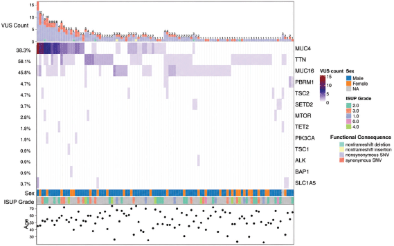
Figure 1. Oncoprint visualisation representing VUS genes and their variants.
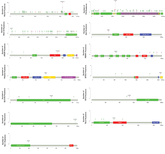
Figure 2. Lollipop graphs of VUS variants identified in the entire cohort.
Statistical analysis
Demographic and baseline clinical characteristics are described using descriptive statistics. Comparisons of different features between patients with at least one VUS variant and those without are performed using different statistical tests, such as chi-square, Fisher’s exact, Mann–Whitney U and chi-square for trend. Univariable and multivariable logistic regression analyses were performed to identify factors associated with the presence of VUS. VUS status was considered the dependent variable, and results are reported as odds ratios with 95% confidence intervals. The multivariable model including age, sex, histology and IMDC risk group was considered the primary model, while alternative multivariable models were evaluated as sensitivity analyses. Given the relatively small number of patients without VUS and the presence of sparse data in some categories, Firth’s penalised logistic regression was additionally performed to reduce potential small-sample bias and assess the robustness of the estimates. A p-value < 0.05 was considered statistically significant in all tests. Statistical analysis was done using R, version 4.5.1.
Results
Baseline characteristics
A total of 139 patients were included in the present analysis. Germline VUS were identified in 108 patients, corresponding to a prevalence of 77.7% in the cohort. The prevalence of VUS differed significantly by sex, with a higher proportion among female patients than among males (93% versus 75%; p = 0.044). The median age of patients harbouring VUS was comparable to that of patients without VUS (54 years [IQR 47–63.75] versus 55 years [IQR 49–65.5]; p = 0.36).
Clear cell RCC constituted the majority of cases (70.5%), with non-clear cell histologies accounting for 24.5%; histology was unavailable for 7 patients (5.0%). Among VUS-positive patients, 69.4% had clear cell histology. The distribution of histological subtypes did not differ significantly between patients with and without VUS (p = 0.774). Similarly, Fuhrman/ISUP grade distribution was comparable between groups, with grade 2 tumours observed in 42.6%, grade 3 in 29.8% and grade 4 in 14.9% of VUS-positive patients (p = 0.303). Metastatic burden, sites of metastasis and IMDC risk categories were also similar between VUS-positive and VUS-negative groups. Detailed baseline characteristics are provided in Table 1.
Univariable and multivariable logistic regression analyses were performed to identify factors associated with VUS status. Univariable logistic regression analysis identified male sex as the only factor significantly associated with VUS status (OR, 0.20; 95% CI, 0.05–0.91; p = 0.038). In the primary multivariable model adjusting for age, sex, histology and IMDC risk group, male sex remained significantly associated with lower odds of having a VUS (OR, 0.11; 95% CI, 0.01–0.88; p = 0.037). Similar findings were observed in the sensitivity analyses (Supplementary Tables 1 and 2).
Spectrum of variants of uncertain significance
A total of 246 germline VUS were identified among 108 of 139 patients (77.7%). The majority of VUS were detected in TTN, MUC16 and MUC4. Following exclusion of these genes, 30 VUS remained, identified in 26 patients (18.7% of the overall cohort).
Among the established RCC susceptibility genes evaluated, VUS were identified in seven patients, with TSC2 being the most frequently involved gene (n = 5), followed by TSC1 (n = 1) and BAP1 (n = 1) (Table 2). No VUS were detected in VHL, FH, PTEN or TP53. Most susceptibility gene VUS were observed in patients with clear-cell RCC.
Among other RCC-associated and cancer-related genes, VUS were most frequently identified in PBRM1 (n = 5), SETD2 (n = 4), SLC1A5 (n = 4), MTOR (n = 3), TET2 (n = 2) and PIK3CA (n = 2), with isolated variants detected in ALK (Table 2). These variants were predominantly observed in patients with clear-cell RCC, although selected alterations were also identified in non-clear-cell subtypes. No significant association was observed between VUS distribution and metastatic burden.
Assessment of variant burden per individual demonstrated that 41 patients harboured a single VUS, 21 patients carried two variants, 17 carried three variants and 29 patients harboured atleast four VUS, with a maximum of 17 variants identified in a single patient. The median number of VUS per patient is 2 (range, 1–19). A total of 20 patients having at least one VUS in the study cohort had metastatic involvement at more than or equal to four sites. 11 of these patients harboured VUS in at least one of the three genes MUC4, MUC16 and TTN (Table 3). The detailed distribution of VUS across individual patients and corresponding gene involvement is summarised in Supplementary Table 3.
Among 246 VUS, 70 putative novel variants without rsIDs were identified. Most new variants were found in the TTN (n = 22) and MUC4 (n = 22), followed by MUC16 (n = 13). Detailed variant annotations of these novel variants are mentioned in Supplementary Table 4.
We further visualised the distribution of all the identified VUS on the gene locations to understand the variant clustering (Figure 2). Compared with the reported variant distribution in ClinVar, no VUS hotspot was identified in the lollipop plots for any of the genes in our cohort.
Population-specific frequency-based reclassification of VUS leveraging IndiGenomes
The VUS identified through ACMG classification were further reclassified using the Indian population allele frequency and zygosity data from the IndiGenomes project. Of the 246 VUS initially identified by ACMG classification, 64 variants (26%) were found in the IndiGenomes database. Notably, three variants within the MUC4 gene (rs12487058, rs529650710 and rs368202391) were reclassified: rs12487058 as benign and rs529650710 and rs368202391 as likely benign. The remaining variants, including those in cancer susceptibility genes, retained their VUS status (Supplementary Table 5).
Table 1. Baseline characteristics.
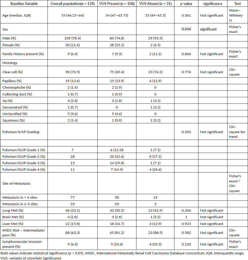
Table 2 . VUS distribution by histological subtype in RCC predisposition and RCC-related genes.
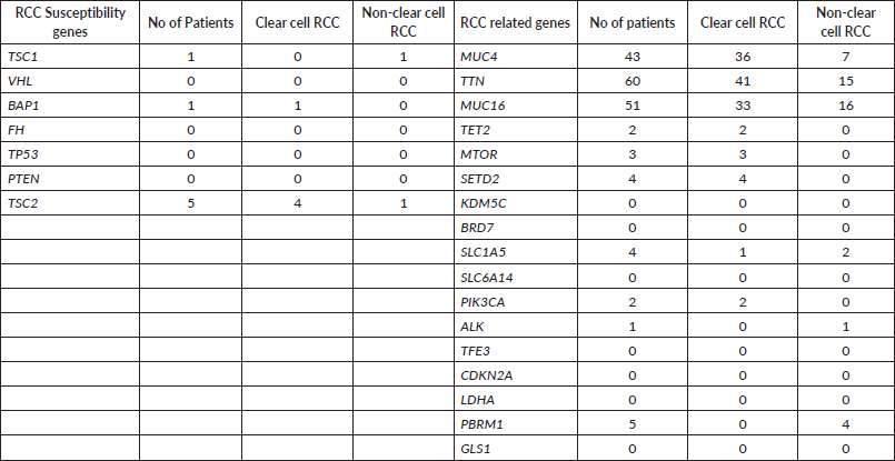
Table 3. Frequency of patients with MUC16, MUC4 and TTN VUS according to metastatic burden.

Cohort-wise allele frequency comparison of the variants
We compared allele frequencies of the variants between the RCC study cohort and publicly available healthy population genomic databases, including IndiGenomes, 1000 Genomes and gnomAD. Novel variants, unique to our cohort, were excluded from this comparison. Five variants (rs12487058, rs150632976, rs199815527, rs201640813 rs529650710) showed statistically significant differences when compared to the IndiGenomes database; of these, all except rs12487058 had higher frequencies in the RCC cohort. Thirteen variants displayed significant allele frequency differences relative to the 1000 Genomes variant database.
For gnomAD, we analysed seven population-specific allele frequencies: South Asian (SAS), East Asian (EAS), Middle Eastern (MID), American (AMR), Ashkenazi Jewish (ASJ), Finnish (FIN) and Non-Finnish European (NFE). Detailed comparisons are visualised in Supplementary Figure 1 using bubble plots. Notably, variant rs112774151 exhibited markedly higher allele frequencies across all seven gnomAD populations (0.216 to 0.318) compared to the RCC cohort (0.0072).
Discussion
This represents the first Indian study evaluating germline VUS across both RCC cancer susceptibility genes and RCC-related oncogenic pathway genes in patients with advanced RCC. Overall, 108 of 139 patients (77.7%) harboured at least one VUS. VUS involving established RCC susceptibility genes were relatively uncommon, being identified in only 7 patients (5.0%), predominantly in TSC2, whereas the majority of variants were detected in genes implicated in RCC pathogenesis and oncogenic signalling pathways. For instance, Carlo et al. [5] reported pathogenic/likely pathogenic germline alterations in 41 of 254 patients (16.1%), including RCC-associated variants in 14 patients (5.5%) and non-RCC cancer predisposition gene alterations in approximately 10.6% (5). Similarly, Wu et al. [28] demonstrated that the use of broader multigene panels increased the detection of germline alterations, highlighting the impact of panel composition on variant identification. In contrast, our study specifically evaluated VUS within RCC susceptibility and RCC-related genes, which may partly explain the relatively low frequency of variants observed in established RCC predisposition genes.
Among cancer-related genes, most VUS were identified in TTN, MUC4 and MUC16, accounting for 216 of 246 (87.8%) variants. Although traditionally considered large structural or passenger genes [29], emerging evidence suggests that alterations in these genes may influence tumour progression, immune modulation and therapeutic responsiveness. TTN mutations have been associated with tumour mutational burden and response to immune checkpoint inhibitors [30,31], while MUC4 and MUC16 are implicated in immune evasion and metastatic dissemination across multiple solid tumours [32]. The high frequency of VUS in these genes highlights the need for further functional characterisation and clinical correlation, particularly given their potential role in shaping tumour behaviour and treatment outcomes. In this context, the observation that more than 50% of the patients having at least one VUS with metastases at more than four sites harboured VUS in at least one of MUC4, MUC16 or TTN is biologically plausible, but must be interpreted with caution in light of the high background mutational burden and sizeable coding regions of these genes, which predispose them to accumulating passenger variants. While this pattern raises a hypothesis that VUS in these loci could contribute to more aggressive or disseminated disease phenotypes, it may also represent a coincidental enrichment rather than a truly causal association, underscoring the need for larger, independently validated cohorts and functional studies. Notably, the significantly higher VUS burden observed among female patients in our cohort is consistent with reports of gender-specific TTN alterations as a potential biomarker of female risk in clear cell RCC [33].
Recent integrative genomic studies further contextualise these findings. Dizman et al. [34] demonstrated that alterations in key RCC-associated genes influence differential response to immunotherapy and targeted therapy. In metastatic RCC, PBRM1 mutations were associated with improved clinical benefit, whereas TERT promoter mutations were mutually exclusive with PBRM1 and associated with lack of benefit from immunotherapy [34]. Furthermore, SETD2 mutations were linked to inferior progression-free and overall survival, suggesting adverse prognostic significance [35]. Several genes in which VUS were identified in our study, including SETD2, KDM5C, TSC2 and MTOR, play established roles in chromatin remodelling, epigenetic regulation and oncogenic signalling pathways that influence tumour aggressiveness and therapeutic response [36]. In addition, the recurrence of VUS in SLC1A5, a glutamine transporter, is consistent with the recognised contribution of metabolic reprogramming to RCC subtypes and clinical behaviour [37]. Notably, PBRM1 VUS were observed at a relatively lower frequency compared to variants in genes associated with aggressive tumour biology. Although the variants identified in our study were classified as uncertain, their occurrence within biologically and clinically relevant pathways and in patients with higher metastatic burden raises the possibility that some may have functional consequences affecting disease behaviour or treatment outcomes. Importantly, because whole-exome sequencing in this study was performed on peripheral blood, the variants reported here represent germline alterations. The prognostic and predictive associations described above for PBRM1, TERT and SETD2 were established for somatic tumour alterations [34,35] and therefore cannot be directly extrapolated to the germline variants of uncertain significance identified in the present cohort; these somatic findings are cited here to provide biological context for the relevance of these genes in RCC rather than to imply equivalent clinical effects for germline VUS.
Variants were also detected in ALK and TFE3, genes increasingly recognised as defining molecular subsets under the WHO 2022 classification of renal tumours [2]. Although classically associated with distinct translocation-driven RCC subtypes, the presence of VUS in these genes within clear cell background histology highlights molecular heterogeneity that may not be fully captured by morphology alone.
In our study, only three VUS were reclassified as benign or likely benign after incorporating Indian-specific allele frequency and zygosity data, which may in part reflect the fact that only 64 of the 246 VUS were represented in the IndiGenomes database. This limited overlap underscores the need for ongoing curation and expansion of Indian genomic reference datasets, as well as periodic re-evaluation of VUS as new evidence becomes available. VUS present substantial clinical challenges, as they neither confirm hereditary cancer risk nor offer clear therapeutic guidance, and prior studies have shown that patients often misunderstand VUS results and can experience psychological distress comparable to that seen with pathogenic variant reports. Therefore, integrating functional assays, multi-omics approaches and longitudinal clinical outcome studies is essential to refine variant classification and ultimately improve the clinical utility of germline and somatic genomic testing in Indian populations [38–40].
To our knowledge, this represents one of the first efforts to develop a germline VUS-focused genomic dataset in Indian patients with advanced RCC. However, several limitations exist. First, treatment outcomes and survival correlations were not evaluated in patients harbouring VUS in cancer-related genes, limiting assessment of their prognostic or predictive significance. Second, variants in non-RCC-associated genes, including DNA repair and other oncogenic pathway genes, were not analysed. Third, although this represents one of the largest Indian RCC germline cohorts to date, the sample size remains modest and warrants validation in larger, multicentre studies. Finally, integration with paired tumour tissue sequencing and transcriptomic profiling, which may have provided better delineation of the functional relevance of these variants, was not performed.
Conclusion
In this cohort of Indian patients with advanced RCC, germline VUS were identified in 77.7% of patients; however, only 5.0% harboured VUS in established RCC susceptibility genes. In contrast, the majority of variants occurred in genes implicated in RCC pathogenesis and oncogenic signalling pathways. These findings highlight a substantial burden of unresolved genomic variation in biologically relevant genes that may influence disease behaviour and therapeutic response. Further functional validation and population-specific genomic studies are needed to define their clinical significance.
Conflicts of interest
All authors declare no potential conflicts of interest.
Funding
Supported by an Institutional Research Grant from AIIMS, New Delhi.
Ethical approval
The study protocol was approved by IEC vide letter number: IEC-573/01.09.2023.
Consent to participate
Informed consent was obtained from all patients.
Availability of data and material
Data regarding this study will be available from the corresponding author on reasonable request.
Authors’ contributions
Authors (AB, MDK, PRK, AP, NP, HB, VH, AS, IG, SK, SM and PT ) contributed to the concept, design and conduct of the study. PRK, AP, NP and MDK did the statistical analysis. PRK, AP, NP, AB and MDK drafted the manuscript, and all authors edited the manuscript.
References
1. Ferlay J, Ervik M, and Lam F, et al (2024) Global Cancer Observatory: Cancer Today (Lyon: International Agency for Research on Cancer) [https://gco.iarc.who.int/today] Date accessed: 23/07/25
2. Goswami PR, Singh G, and Patel T, et al (2024) The WHO 2022 classification of renal Neoplasms (5th Edition): salient Updates Cureus 16(4) e58470 https://doi.org/10.7759/cureus.58470
3. Linehan WM (2012) Genetic basis of kidney cancer: role of genomics for the development of disease-based therapeutics Genome Res 22(11) 2089–2100 https://doi.org/10.1101/gr.131110.111
4. Yanus GA, Kuligina ES, and Imyanitov EN (2024) Hereditary renal cancer syndromes Med Sci 12(1) 12 https://doi.org/10.3390/medsci12010012
5. Carlo MI, Mukherjee S, and Mandelker D, et al (2018) Prevalence of germline mutations in cancer susceptibility genes in patients with advanced renal cell carcinoma JAMA Oncol 1228(9) 1228–1235 https://doi.org/10.1001/jamaoncol.2018.1986
6. Hampel H, Bennett RL, and Buchanan A, et al (2015) A practice guideline from the American College of Medical Genetics and Genomics and the National Society of Genetic Counselors: referral indications for cancer predisposition assessment [Internet] Genet Med Off J Am Coll Med Genet 17(1) 70–87 [https://pubmed.ncbi.nlm.nih.gov/25394175/]
7. Mendhiratta N, Hauver H Jr, and Hatton W, et al (2024) Outcomes of a universal germline screening program in a community urology practice Clin Genet 106(3) 277–283 https://doi.org/10.1111/cge.14541
8. Choudry MM, Durant AM, and Edmonds VS, et al (2024) Germline pathogenic variants identified in patients with genitourinary malignancies undergoing universal testing: a multisite single-institution prospective study J Urol 212(4) 590–599 https://doi.org/10.1097/JU.0000000000004089
9. Samadder NJ, Riegert-Johnson D, and Boardman L, et al (2021) Comparison of Universal Genetic Testing vs Guideline-Directed Targeted Testing for Patients With Hereditary Cancer Syndrome JAMA Oncol 7(2) 230–237 https://doi.org/10.1001/jamaoncol.2020.6252 PMCID: 7600058
10. Tung N and Desai N (2021) Germline genetic testing for women with breast cancer: shifting the paradigm from whom to test to whom NOT to test J Clin Oncol 3415(31) 3415–4318 https://doi.org/10.1200/JCO.21.01761
11. Whitworth PW, Beitsch PD, and Patel R, et al (2022) Clinical Utility of Universal Germline Genetic Testing for Patients With Breast Cancer JAMA Netw Open 5(9) e2232787
12. Richards S, Aziz N, and Bale S, et al (2015) Standards and guidelines for the interpretation of sequence variants: a joint consensus recommendation of the American College of Medical Genetics and Genomics and the Association for Molecular Pathology Genet Med 17(5) 405–423 https://doi.org/10.1038/gim.2015.30 PMID: 25741868 PMCID: 4544753
13. Moghadasi S, Eccles DM, and Devilee P, et al (2016) Classification and clinical management of variants of uncertain significance in high penetrance cancer predisposition genes Hum Mutat 37(4) 331–336 https://doi.org/10.1002/humu.22956
14. Yngvadottir B, Andreou A, and Bassaganyas L, et al (2022) Frequency of pathogenic germline variants in cancer susceptibility genes in 1336 renal cell carcinoma cases Hum Mol Genet 3001(17) 3001 https://doi.org/10.1093/hmg/ddac089
15. Nguyen CB, Knaus C, and Li J, et al (2023) Pathogenic germline mutational landscape in patients with renal cell carcinoma and associated clinicopathologic features JCO Precis Oncol 7 e2300168 https://doi.org/10.1200/PO.23.00168 PMID: 38127826
16. Xing J, Watkins WS, and Hu Y, et al (2010) Genetic diversity in India and the inference of Eurasian population expansion Genome Biol 11(11) R113 https://doi.org/10.1186/gb-2010-11-11-r113 PMID: 21106085 PMCID: 3156952
17. Wills C (2007) Principles of population genetics J Hered 98(4) 382 https://doi.org/10.1093/jhered/esm035
18. Zhang C, Gao Y, and Ning Z, et al (2019) PGG.SNV: understanding the evolutionary and medical implications of human single nucleotide variations in diverse populations Genome Biol 20(1) 215 https://doi.org/10.1186/s13059-019-1838-5 PMID: 31640808 PMCID: 6805450
19. Osler TS, Brandenburg JT, and Schoeman M, et al (2024) Prevalence and reclassification of genetic variants in South African populations with breast cancer Genes Chromosomes Cancer 63(9) e23275 https://doi.org/10.1002/gcc.23275 PMID: 39324485
20. Bajaj A, Senthivel V, and Bhoyar R, et al (2022) 1029 genomes of self-declared healthy individuals from India reveal prevalent and clinically relevant cardiac ion channelopathy variants Hum Genomics 16(1) 30 https://doi.org/10.1186/s40246-022-00402-2 PMID: 35932045 PMCID: 9354277
21. Nagpal C, Divakar MK, and Baskarane H, et al (2025) Prevalence of germline variants in advanced renal cell Carcinoma in North India JCO Glob Oncol 11 e2500137 https://doi.org/10.1200/GO-25-00137
22. Barata PC and Rini BI (2017) Treatment of renal cell carcinoma: current status and future directions CA Cancer J Clin 67(6) 507–524 [https://doi.org/10.3322/caac.21411] PMID: 28961310
23. Lalani AKA, Heng DYC, and Basappa NS, et al (2022) Evolving landscape of first-line combination therapy in advanced renal cancer: a systematic review Ther Adv Med Oncol 14 17588359221108685
24. Moch H (2016) The WHO/ISUP grading system for renal carcinoma Pathol 37(4) 355–360 https://doi.org/10.1007/s00292-016-0171-y
25. Fuhrman SA, Lasky LC, and Limas C (1982) Prognostic significance of morphologic parameters in renal cell carcinoma Am J Surg Pathol 6(7) 655–663 https://doi.org/10.1097/00000478-198210000-00007 PMID: 7180965
26. Xie D, Li G, and Zheng Z, et al (2025) The molecular code of kidney cancer: a path of discovery for gene mutation and precision therapy Mol Aspects Med 101 101335
27. Jain A, Bhoyar RC, and Pandhare K, et al (2021) IndiGenomes: a comprehensive resource of genetic variants from over 1000 Indian genomes Nucleic Acids Res 49(D1) D1225–D1232
28. Wu J, Wang H, and Ricketts CJ, et al (2019) Germline mutations of renal cancer predisposition genes and clinical relevance in Chinese patients with sporadic, early-onset disease Cancer 1060(7) 1060–1109 https://doi.org/10.1002/cncr.31908
29. Tharp CA, Haywood ME, and Sbaizero O, et al (2019) The Giant Protein Titin’s Role in Cardiomyopathy: genetic, Transcriptional, and Post-translational Modifications of TTN and Their Contribution to Cardiac Disease Front Physiol 1436 1436 https://doi.org/10.3389/fphys.2019.01436
30. Jia Q, Wang J, and He N, et al (2019) Titin mutation associated with responsiveness to checkpoint blockades in solid tumors JCI Insight 4(10) e127901 [https://pubmed.ncbi.nlm.nih.gov/31092729/] https://doi.org/10.1172/jci.insight.127901
31. Yang Y, Zhang J, and Chen Y, et al (2020) MUC4, MUC16, and TTN genes mutation correlated with prognosis, and predicted tumor mutation burden and immunotherapy efficacy in gastric cancer and pan‐cancer Clin Transl Med 10(4) e155 https://doi.org/10.1002/ctm2.155 PMID: 32898332 PMCID: 7443139
32. Bhatia R, Gautam SK, and Cannon A, et al (2019) Cancer-associated mucins: role in immune modulation and metastasis Cancer Metastasis Rev 38(1-2) 223–236 [https://pubmed.ncbi.nlm.nih.gov/30618016/] https://doi.org/10.1007/s10555-018-09775-0
33. Saha A, Sharmin S, and Ahmed T, et al (2025) Gender-specific insights into TTN mutation: potential biomarker for female risk in kidney renal clear cell carcinoma J Genet Eng Biotechnol 23(3) 100506 https://doi.org/10.1016/j.jgeb.2025.100506 PMID: 40854626 PMCID: 12169730
34. Dizman N, Lyou Y, and Salgia N, et al (2020) Correlates of clinical benefit from immunotherapy and targeted therapy in metastatic renal cell carcinoma J Immunother Cancer 8(2) e000953 [https://pubmed.ncbi.nlm.nih.gov/32661119/] https://doi.org/10.1136/jitc-2020-000953
35. Wang J, Liu L, and Qu Y, et al (2016) Prognostic value of SETD2 expression in patients with metastatic renal cell carcinoma treated with tyrosine kinase inhibitors J Urol 196(5) 1363–1370 [https://pubmed.ncbi.nlm.nih.gov/27288695/] https://doi.org/10.1016/j.juro.2016.06.010
36. Piva F, Santoni M, and Matrana MR, et al (2015) BAP1, PBRM1 and SETD2 in clear-cell renal cell carcinoma: molecular diagnostics and possible targets for personalized therapies Expert Rev Mol Diag 15(9) 1201–1210 [https://pubmed.ncbi.nlm.nih.gov/26166446/] https://doi.org/10.1586/14737159.2015.1068122
37. Yao Z, Zheng Z, and Zheng X, et al (2022) Comprehensive characterization of metabolism‐associated subtypes of renal cell carcinoma to aid clinical therapy Oxid Med Cell Longev2022(1) 9039732 https://doi.org/10.1155/2022/9039732
38. Walsh N, Cooper A, and Dockery A, et al (2024) Variant reclassification and clinical implications J Med Genet 61(3) 207–211 https://doi.org/10.1136/jmg-2023-109488 PMID: 38296635
39. Mighton C, Shickh S, and Uleryk E, et al (2021) Clinical and psychological outcomes of receiving a variant of uncertain significance from multigene panel testing or genomic sequencing: a systematic review and meta-analysis Genet Med 23(1) 22–33 https://doi.org/10.1038/s41436-020-00957-2
40. Makhnoon S, Garrett LT, and Burke W, et al (2019) Experiences of patients seeking to participate in variant of uncertain significance reclassification research J Community Genet 10(2) 189–196 https://doi.org/10.1007/s12687-018-0375-3 PMCID: 6435771
Supplementary information
Supplementary information can be viewed at https://doi.org/10.5281/zenodo.23186801